MATERIALS AND METHODS
Case Material
The cases analyzed for this study consisted of 447 GIST, 935 other mesenchymal tumors (Table 1), and 432 non-sarcomatous tumors distributed over 10 TMAs. The TMAs were constructed using a manual tissue arrayer from Beecher Instrument, Silver Spring, MD. Cores of 0.6 mm were taken from paraffin embedded soft tissue tumors from the Stanford University Medical Center, the Oregon Health & Science University, Portland, Oregon, and the University of British Columbia, Vancouver. Mutational information was available for 306 of the 447 GIST cases. The GIST were analyzed for mutations in exons 9, 11, 13, and 17 of the KIT gene using a combination of denaturing high pressure liquid chromography and direct sequencing, as previously described. 3, 9 KIT wild-type tumors were subsequently screened for mutations in exons 12, 14, and 18 of the PDGFRA gene.
Immunohistochemistry
Slides were cut at 4μm, deparaffinized in xylene and hydrated in a graded series of alcohol. The primary antibodies used were DOG 1.1 (mouse monoclonal, 1/50; Stanford University.), DOG1.3 (mouse monoclonal, 1/1 supernatant; Applied Genomics Inc.), CD117 (rabbit polyclonal, 1/400; DAKO, Carpinteria, CA), and CD34 (mouse monoclonal, 1/80; clone 581/CD34, BD Biosciences, San Jose, CA). The antigen retrieval solution for DOG1.1, DOG1.3 and CD34 was citrate, pH: 6 and for CD117 was EDTA, pH:8. Slides were boiled by microwaving in antigen retrieval solution for 12 minutes. The immunohistochemical reactions were visualized using rabbit or mouse versions of the EnVision + system (DAKO, Carpinteria, CA) using diaminobenzidine.
In Situ Hybridization
In situ hybridization of TMA sections was performed based on a protocol published previously.
Scoring of immunohistochemistry and in situ hybridization
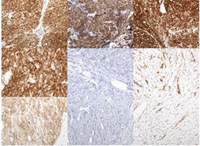
Cores were scored as follows: 0 indicates the absence of any staining; 1 indicates equivocal staining; 2 indicates any moderate membranous staining whether diffusely or focally present in the tumor; 3 indicates strong complete membranous staining whether diffusely or focally present in the tumor (Fig. 1A-D). Score 2 and 3 were considered positive. The cores were independently reviewed for two pathologists (IE and CHL) and any disagreements were reviewed together with a third pathologist (MvdR) to achieve a consensus score.
Antibody generation
The peptides sequences to generate the DOG1.1 and DOG1.3 monoclonal antibodies were MSDFVDWVIPDIPKDISQQIHKEKVLMVELFMREEQDKQQLLETCMEKER
QKDEPPCNHHNTKACPDSLGSPAPSHAYHGGVL and KVDYILVYHH
KRPSGNRTLVRRVQHSDTPSGARSVKQDHPLPGKGASLDAGSGEPPMDYHEDDKRFRREEYEGNLLEAGLELERDEDTKIHGVGFVKIHAP, respectively. These peptides were injected intraperitoneally with Freund complete adjuvant into a mouse and boosted 4 times weekly. Spleens were taken for hybridoma preparation as described by Köhler and Milstein.
The fused cells were distributed in 96-well plates and the antibody producing wells were detected by ELISA using DOG1 peptide coated plates. Positive wells were next screened by immunohistochemistry on a mini-TMA containing 5 cases of GIST and 2 cases of leiomyosarcoma.
|