25
|
|
|
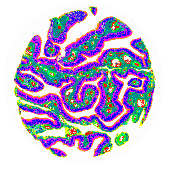
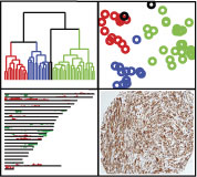
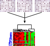
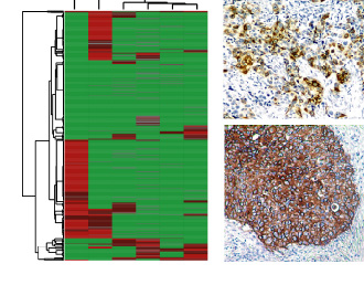
Please see the web supplement for interactive access to images of all stained cores and associated visualizations.
Riordan DP, Varma S, West RB, Brown PO (2015) Automated Analysis and Classification of Histological Tissue Features by Multi-Dimensional Microscopic Molecular Profiling. PLoS ONE 10(7): e0128975. doi:10.1371/journal.pone.0128975
|
24
|
|
|
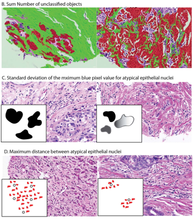
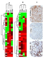
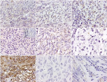
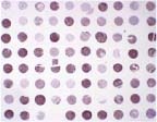
Please see the web supplement for interactive access to images of all stained cores and associated figures.
AH Beck et al., Systematic Analysis of Breast Cancer Morphology Uncovers Stromal Features Associated with Survival, Sci. Transl. Med. 3, 108ra113 (2011).
|
23
|
|
|
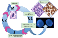
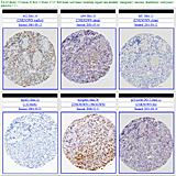
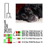
Please see the web supplement for interactive access to images of all stained cores and associated figures.
Modern Pathology advance online publication 8 January 2010; doi: 10.1038/modpathol.2009.173
|
22
|
|
|
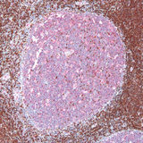
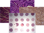
Please see the web supplement for interactive access to images of all stained cores.
Human
Pathology, epub ahead of print (PMID: 20004001)
|
21
|
|
|
Please see the web supplement for interactive access to images of all stained cores and fgures.
Oncogene, doi: 10.1038/onc.2009.381
|
20
|
|
|
Please see the web supplement for interactive access to images of all stained cores and figures.
American Journal of Pathology, doi:10.2353/ajpath.2009.081037
|
19
|
|
|
Please see the web supplement for interactive access to images of all stained cores, figures and supplementary data.
Clinical Cancer Research 15, 778-787, February 1, 2009. doi: 10.1158/1078-0432.CCR-08-1283
|
18
|
|
|
Please see the web supplement for interactive access to a cluster heatmap and images of all stained cores.
The American Journal of Surgical Pathology, August 14, 2008.
|
17
|
|
|
Please see the web supplement for figures, interactive access to images of all stained cores and supplemental data.
American Journal of Clinical Pathology Volume 130, Number 3 / September 2008..
|
16
|
|
|
Please see the web supplement for figures, interactive access to images of all stained cores and supplemental data.
Clinical Cancer Research 14, 1423-1430, March 1, 2008.
|
15
|
|
|
Please see the web supplement for interactive access to images of all stained
cores as well as figures, methods and tables.
American Journal of Surgical Pathology, February 2008, 32:2
|
14
|
|
|
Please see the web supplement for interactive access to images of all stained cores as well as figures and tables.
American Journal of Surgical Pathology, May 2007, 31:5
|
13
|
|
|
Please see the web supplement for interactive access to
supplementary images.
Am J Pathol. 2007 Apr;170(4):1362-9.
|
12
|
|
|
Please see the web supplement for interactive access to
supplementary images.
haematologica 2007;91:176-183
|
11
|
|
|
Visit the web supplement for interactive access to
supplementary images.
Blood First Edition Paper, prepublished online October 12, 2006
|
10
|
|
|
Visit the web supplement for interactive access to the
raw data, full resolution downloadable images and supplemental
material from the authors.
Use GeneXplorer and CaseXplorer to interactively browse the
Gene Microarray and Tissue Microarray data, including stained tissue
cores.
PNAS published January 6, 2006, 10.1073/pnas.0507321103
(Medical Sciences), an Open Access article.
|
9
|
|
|
This site describes the recently released TMA-Combiner, a software tool
used in some of the projects described in this portal. This tool enables
the TMA user to combine multiple replicates from one or more separate TMAs
into a single dataset, with three score combination rules available to
choose from. This tool is also described in detail at Modern
Pathology, doi:10.1038/modpathol.3800491.
|
8
|
|
|
Visit the web supplement for interactive access to
the raw data and downloadable versions of all figures from the paper.
In addition the authors provide supplementary data and images.
The paper was published May, 2005 in Journal of Pathology 206. DOI: 10.1002/path.1792
|
7
|
|
|
Visit the web supplement for interactive access to
supplementary images.
This paper is a Blood First Edition Paper, prepublished online DOI 10.1182/blood-2004-08-3112.
|
6
|
|
|
Visit the web supplement for interactive access to raw data,
figures and tables in the paper and supplementary
figures and tables.
The
paper was published in 2004 in Oncogene 23(47),
pp 7780-9.
|
5
|
|
|
The
web supplement for this paper includes interactive access to
the raw data and downloadable versions of all figures from the paper.
The
paper was published in 2004 in Clinical Cancer Research Vol. 10, pp 5367-5374.
|
4
|
|
|
The web supplement for this paper provides access to an
interactive method to explore the clustered TMA data. The
paper was published in 2004 in the American Journal of
Surgical Pathology, Vol. 28, pp 1063-1069.
|
3
|
|
|
This site provides access to the figures from the paper and an
interactive method to explore the clustered TMA data. The
paper was published in 2004 in the American Journal of
Pathology, Vol. 165, pp 107-113.
|
2
|
|
|
Visit the web supplement to Nielsen et al., Tissue microarray validation of epidermal
growth factor receptor and SALL2 in synovial sarcoma with comparison
to tumors of similar histology. American Journal of
Pathology, Vol. 163, No. 4, pp. 1449-1456.
|
1
|
|
|
This site describes the software tools used in the projects
described in this portal. American Journal of
Pathology, Vol. 161, No. 5, pp. 1557-1565.
|