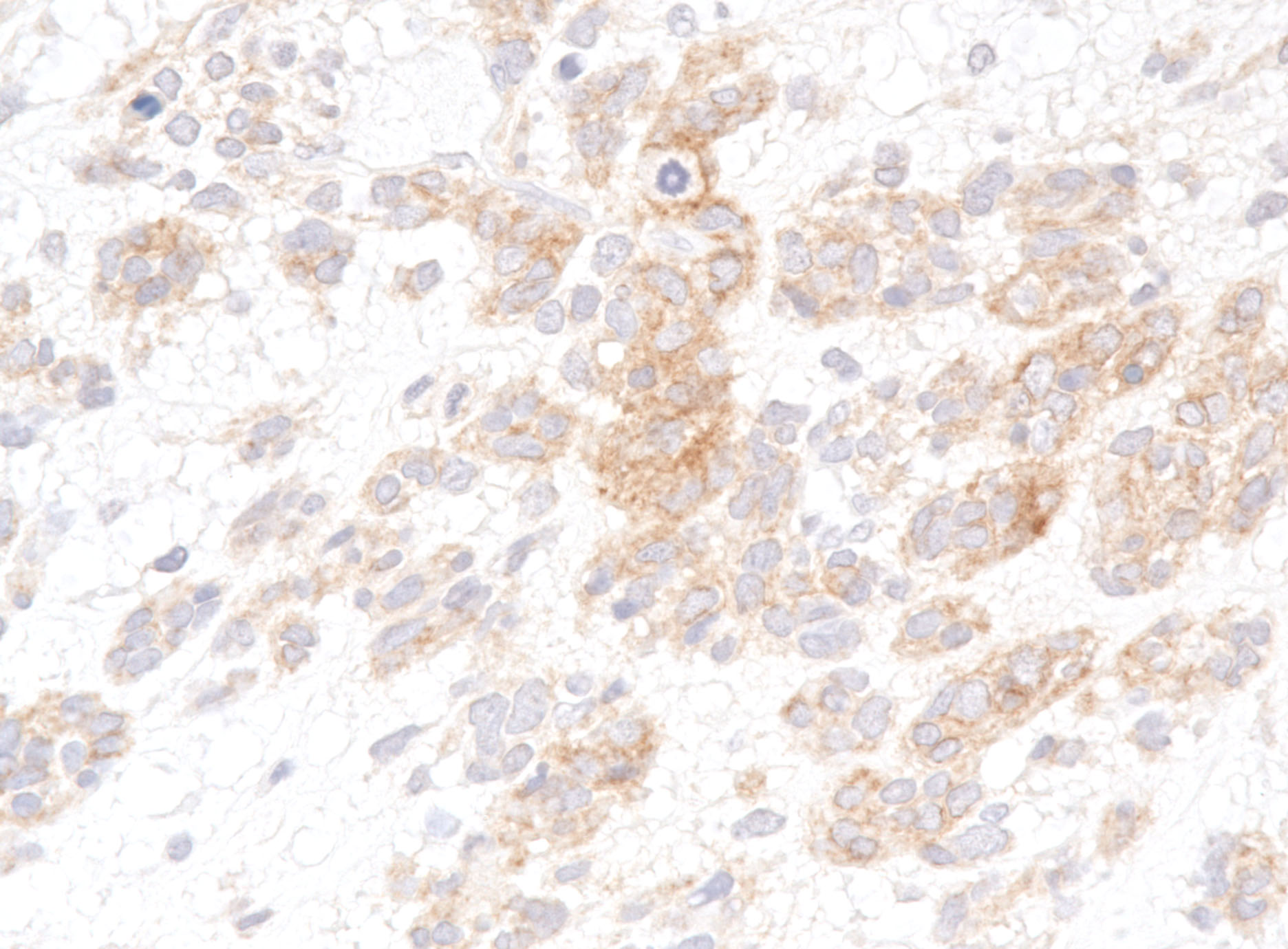
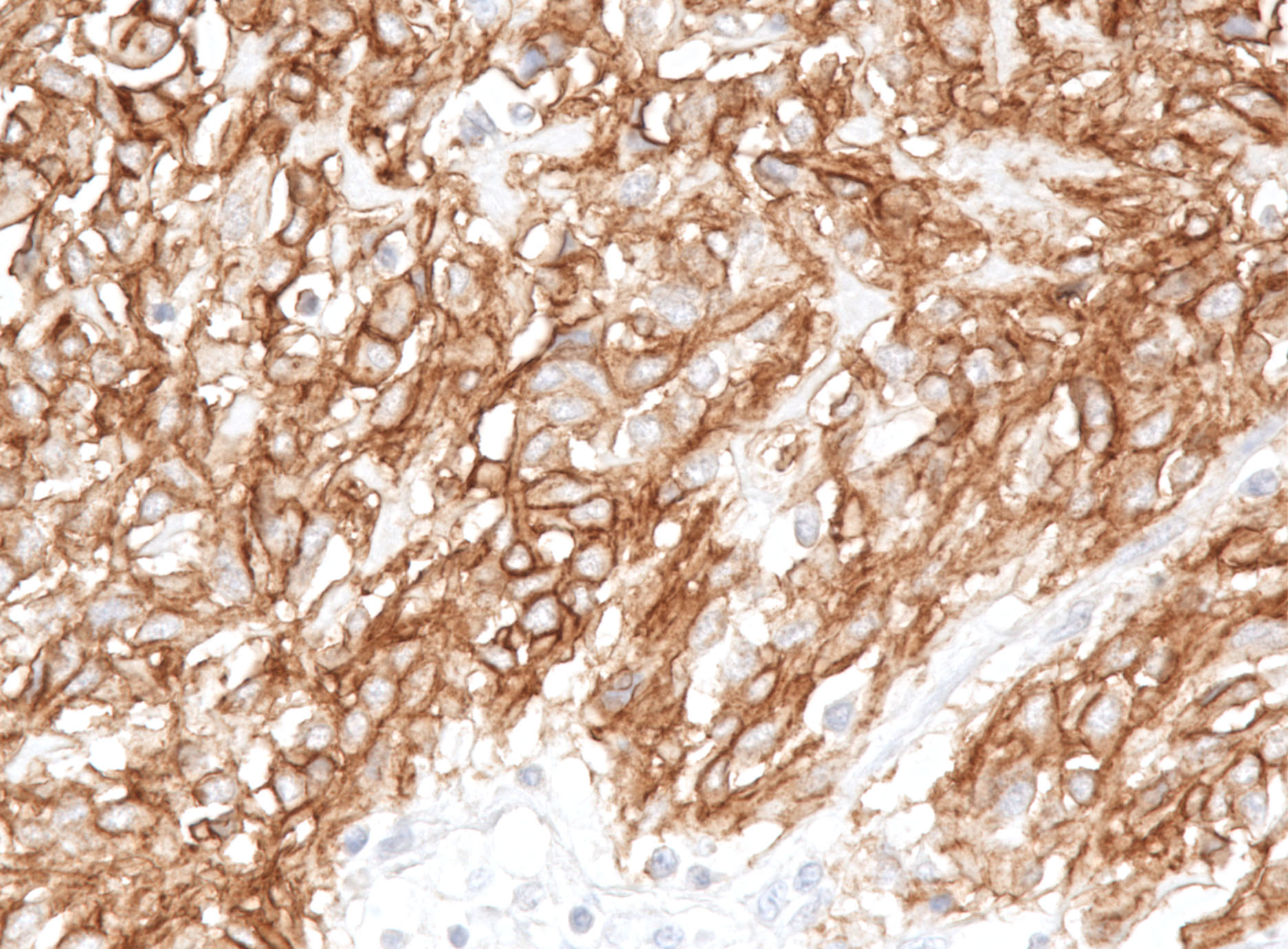
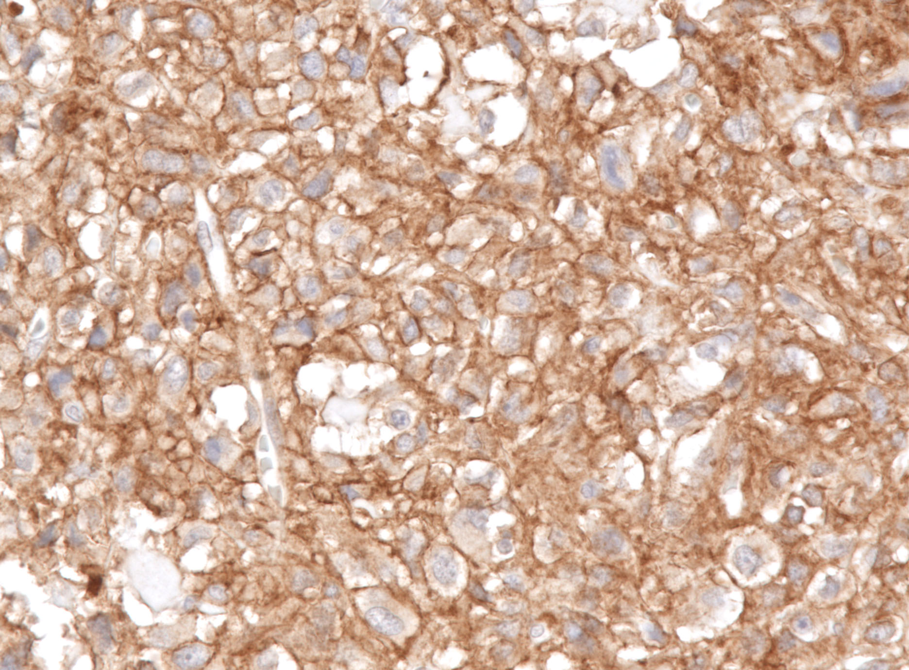
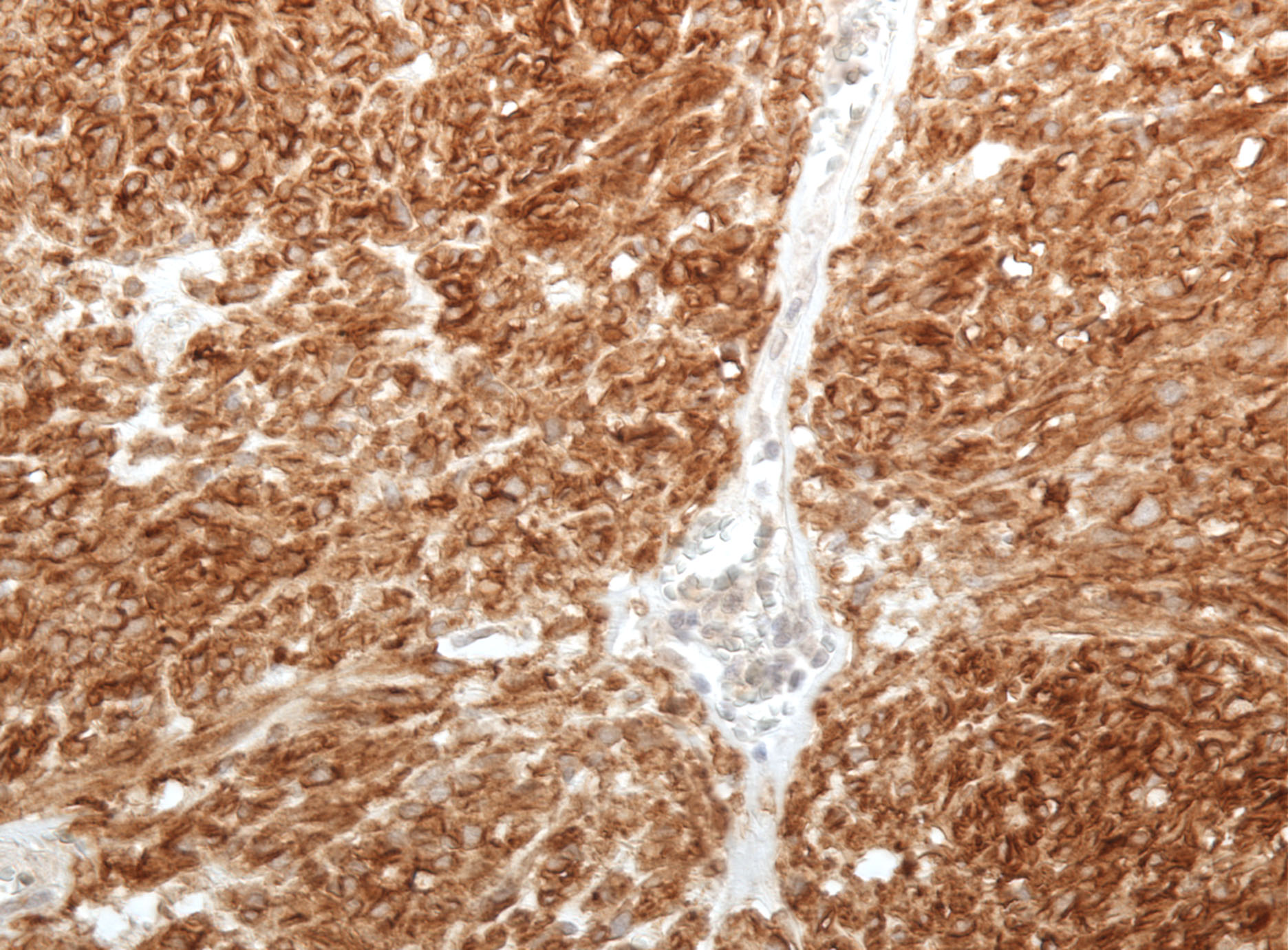
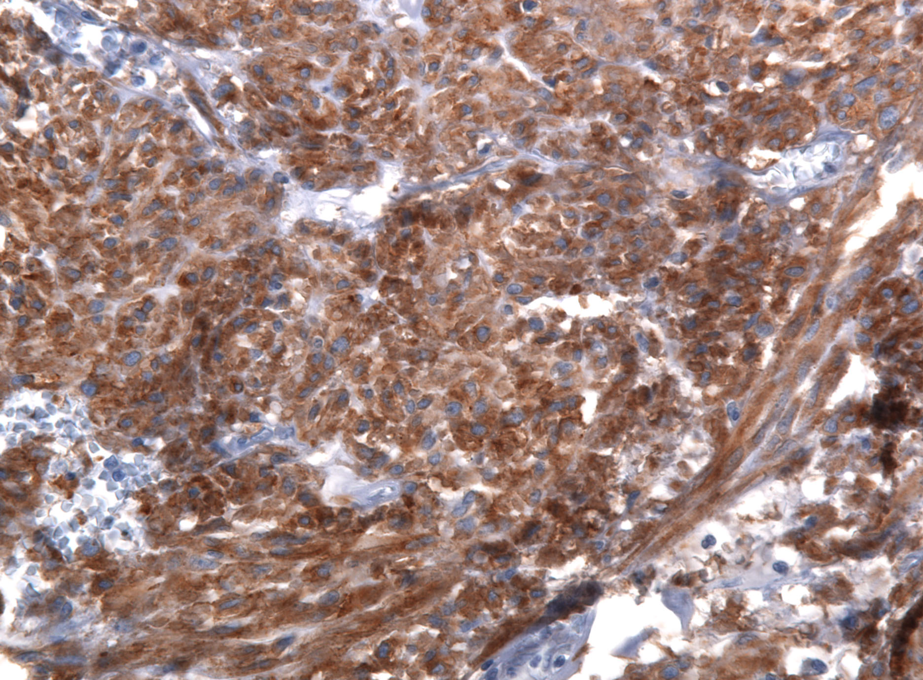
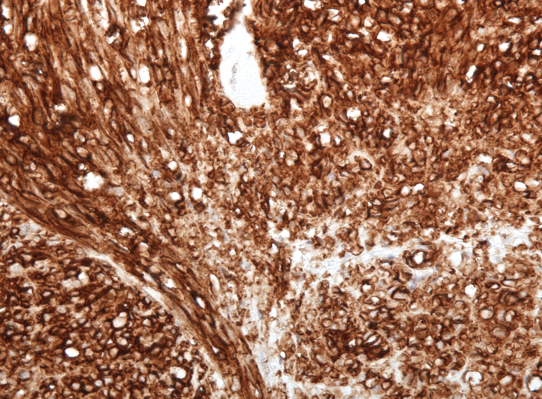
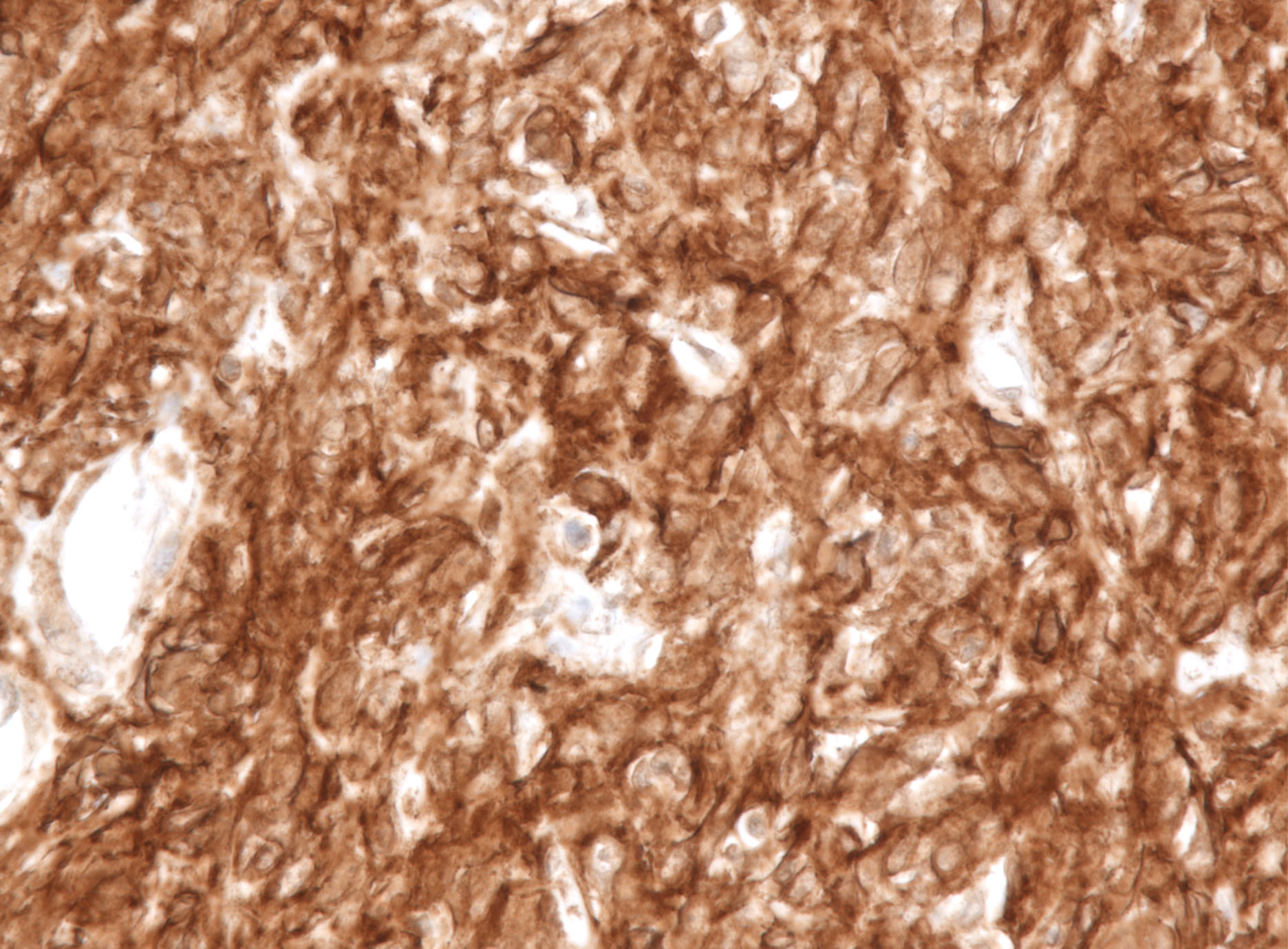
A novel monoclonal antibody against DOG1 is a sensitive and specific marker for gastrointestinal stromal tumors.

Inigo Espinosa, MD a, Cheng-Han Lee, MD, PhDa, Mi Kyung Kim, MD, PhD a, Bich-Tien Rouse, MSa, Subbaya Subramanian, PhD a, Kelli Montgomery a, Sushama Varma, MS a Christopher L. Corless, MD, PhD b, Michael C. Heinrich, MD b, Kevin S. Smith, PhDa, Zhong Wang, PhDa, Brian Rubin MD, PhDc, Torsten O Nielsen MD, PhDd, Robert S Seitz MDe, Douglas T Ross, MD, PhDe , Robert B West, MD, PhDa, Michael L. Cleary, MDa, and Matt van de Rijn, MD, PhDa


Home
Home
Figures
Figures
Images
Images
Tables
Tables
Methods
Methods
Authors
Authors
Web Portal
Web portal

Figures

Figure 1

DOG1.1 immunohistochemistry in GIST (X40). A, Score 0. B, Score 1. C,

Score 2. D, Score 3. E, Strong membranous staining in an epithelioid GIST. F, Strong

cytoplasmic staining in a spindle cell GIST.

A                                                             B                                                            C

D                                                              E                                                             F

Figure 2

Immunohistochemical results for KIT, CD34, and DOG1.1 in KIT or PDGFRA

mutation positive and KIT/PDGFRA mutation negative (WT) GISTs.

Figure 3

Patterns of DOG1.1, KIT, and CD34 in GISTs with different genotypes (X40).

A, KIT exon 11 mutant with expression of DOG1.1 (A1), KIT (A2), and CD34 (A3).

B, KIT exon 11 mutant with expression of DOG1.1 (B1), and negative for KIT (B2)

and CD34 (B3).C, PDGFRa exon 18 mutant with expression of DOG1.1

(C1) and negative for KIT (C2) and CD34 (C3).

A1                                                           A2                                                          A3

B1                                                          B2                                                           B3

C1                                                          C2                                                            C3


Figure 4

DOG1.1 (A) and KIT (B) show similar staining patterns consistent with ICC staining in

the small bowel.

A

B


                      [ Home | Figures | Images | Tables | Methods | WebPortal | Authors]