|
The TMA data management system described here was designed
with the Bliss filename nomenclature in mind. If you created and
curate your digital image collection with the Bliss microscope system,
your files will already be in this format. If you have not, the
following will describe how the nomenclature is structured so that you
can adapt your system appropriately.
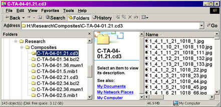
Images generated by the Bliss microscope system (Bacus Labs Inc.) are
stored in the following directory structure:

Click for a larger view
All TMA directories should be contained in the directory
"Composites", as shown above. Each directory or folder represents a collection of images
from a single sector of a given slice of the TMA, as shown above. The total number of
folders in the selection will equal the product of the number of
arrays, slices, and sectors in your TMA collection. In the example
above, which also represents the files stored in the on-line
demonstration database, there are 8 folders, representing an array
composed of 4 slices and 2 sectors each.
The directory name is composed of the
following:
C-TA-0x-0y.z.Ab-Name
An example of this would be the following:
C-TA-04-02.34.bcl2
where:
| Variable |
Description |
Example |
| x |
TMA Array Number |
4 |
| y |
TMA Sector |
2 |
| z |
TMA Slice Number |
34 |
| Ab-Name |
Antibody Name |
bcl2 |
So, in the above example, the folder/directory represents
images collected from TMA number 4, sector 2, slice 34, stained with the antibody against bcl2.
Each of the image filenames in these directories consist of the following:
s_a_c_r_sl_code_d.jpg
An example of this would be the following:
1_4_1_1_34_1012_1.jpg
where:
| Variable |
Description |
Example |
| s |
Sector Number |
1 |
| a |
Array Number |
4 |
| c |
Column Number |
1 |
| r |
Row Number |
1 |
| sl |
Slice Number* |
34 |
| code |
Bliss Code Number* |
1012 |
| d |
Order Number |
1 |
* Note that the raw scoring workbooks
used to generate the TMA TreeView files must contain the necessary
information in order for
Stainfinder to recognize the filenames. This is why the TMA
Deconvoluter will warn the user if "Bliss or slice information" is
missing from their scoring worksheets.
So, in the above example, the folder/directory represents
image collected from Sector 1, Array 4, Column 1, Row 1, Slice 34,
Bliss Code designation 1012, and is the first picture scanned in the
array.
A note about the Bliss code numbers: The BLISS code number
is a numerical code for the name of the antibody used in staining and
is required since the name of a JPG image file generated by the Bliss system
consists of 7 numbers separated by underscores, but does not allow for
letters.
Your digital image collection must follow this
nomenclature in order for Stainfinder to work properly. If there are
missing or incorrect information, Stainfinder may not be able to
retrieve the image file or may retrieve incorrect image files. Please
examine the on-line demonstration database and accompanying files from
the Downloads section to see how a properly configured system should behave.
Note: The Bliss microscope system employs a serpentine scanning
method in generating the order of pictures collected. The Order
Numbers reflect this scanning method. An example of a serpentine scan
is shown below, where the numbers represent the order in which the
images are acquired.
Serpentine Scan
| Sector n |
Column |
| Row |
1 |
2 |
3 |
4 |
5 |
6 |
7 |
8 |
9 |
10 |
11 |
| 1 |
1 |
2 |
3 |
4 |
5 |
6 |
7 |
8 |
9 |
10 |
11 |
| 2 |
22 |
21 |
20 |
19 |
18 |
17 |
16 |
15 |
14 |
13 |
12 |
| 3 |
23 |
24 |
25 |
26 |
27 |
28 |
29 |
30 |
31 |
32 |
33 |
|
|
etc.... |
Scanning beings at Row 1, Column 1 of Sector n and proceeds
to the end of the row. It then moves one row down and proceeds to the
left, until it reaches the left end of the row. It continues onto the
third row and proceeds in like fashion until the end of the sector is reached.
back to top
Return to the Stainfinder walkthrough.
|

