Amy Heerema-McKenney, Liliane CD Wijnaendts, Joseph F Pulliam, Dolores Lopez-Terrada, Jesse K McKenney, Shirley Zhu, Kelli Montgomery, Janet Mitchell, Robert J Marinelli, Augustinus A.M. Hart, Matt van de Rijn, Sabine C Linn
| Home |
|
Home |
| Images |
|
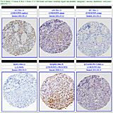
View the rhabdomyosarcoma tissue array images |
| Data |
|
View the rhabdomyosarcoma data |
| WebPortal |
|
Stanford Tissue Microarray Consortium Web Portal |
| Authors |
|
Authors |
| Home |
Welcome to the web supplement to the paper:Diffuse Myogenin Expression by Immunohistochemistry is an Independent Marker of Poor Survival in Pediatric Rhabdomyosarcoma: A Tissue Microarray Study of 71 Primary Tumors Including Correlation With Molecular Phenotype Published August 4, 2008 in The American Journal of Surgical Pathology, Publish Ahead of Print Abstract:The pathologic classification of rhabdomyosarcoma (RMS) into embryonal or alveolar subtype is an important prognostic factor guiding the therapeutic protocol chosen for an individual patient. Unfortunately, this classification is not always straightforward, and the diagnostic criteria are controversial in a subset of cases. Ancillary studies are used to aid in the classification, but their potential use as independent prognostic factors is rarely studied. The aim of this study is to identify immunohistochemical markers of potential prognostic significance in pediatric RMS and to correlate their expression with PAX-3/FKHR and PAX-7/FKHR fusion status. A single tissue microarray containing 71 paraffin-embedded pediatric RMSs was immunostained with antibodies against p53, bcl-2, Ki-67, CD44, myogenin, and MyoD1. The tissue microarray and whole paraffin blocks were studied for PAX-3/FKHR and PAX-7/FKHR gene fusions by fluorescence in situ hybridization and reverse transcription-polymerase chain reaction. Clinical follow-up data were available for each patient. Immunohistochemical staining results and translocation status were correlated with recurrence-free interval (RFI) and overall survival (OS) using the Kaplan-Meier method, the log-rank test, and Cox proportional hazard regression. The minimum clinical follow-up interval was 24 months (median follow-up=57 mo). On univariable analysis, immunohistochemical expression of myogenin, bcl-2, and identification of a gene fusion were associated with decreased 5-year RFI and 10-year OS (myogenin RFI P=0.0028, OS P=0.0021; bcl-2 RFI P=0.037, OS P=0.032; gene fusion RFI P=0.0001, OS P=0.0058). After adjustment for Intergroup Rhabdomyosarcoma Study-TNM stage, tumor site, age, tumor histology, and translocation status by multivariable analysis, only myogenin retained an independent association with RFI (P=0.034) and OS (P=0.0069). In this retrospective analysis, diffuse immunohistochemical reactivity for myogenin in RMS correlates with decreased RFI and OS, independent of histologic subtype, translocation status, tumor site, or stage. |