Prognostic significance of macrophage infiltration in leiomyosarcomas
Cheng-Han Lee1, 2 †, Inigo Espinosa1†, SuzanVrijaldenhoven1,
Subbaya Subramanian1,Kelli D Montgomery1, Shirley Zhu1, Robert J Marinelli3, Johannes L Peterse 4, Neal Poulin2, Torsten O. Nielsen2, Rob B. West1,C. Blake Gilks2, Matt van de Rijn1.
Home
|
Figures and Tables |
View the tissue
array images |
Supplemental data |
| Authors |
Authors
|
| Supplemental data |
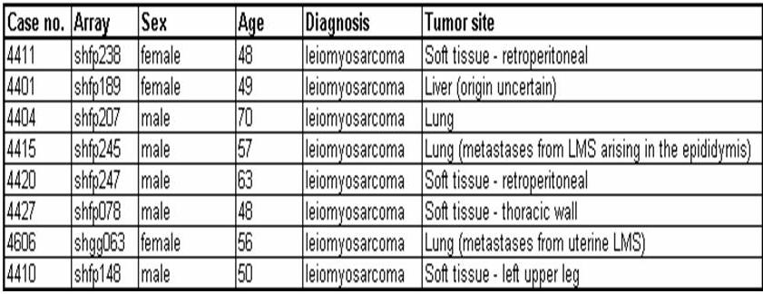
Web supplement Table 1: Clinicopathologic features of the fresh frozen LMS samples used for gene array analysis.
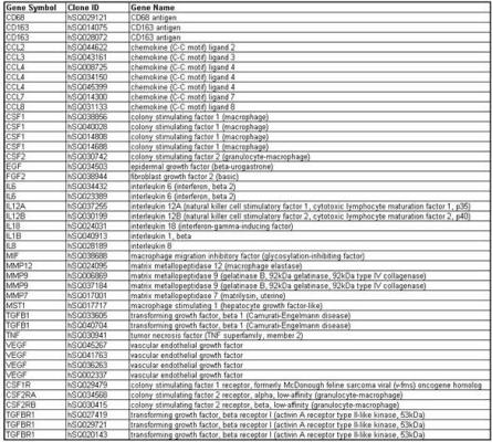
Gene list for gene array analysis
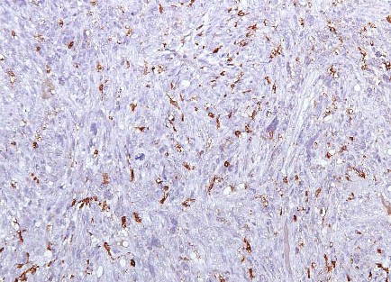
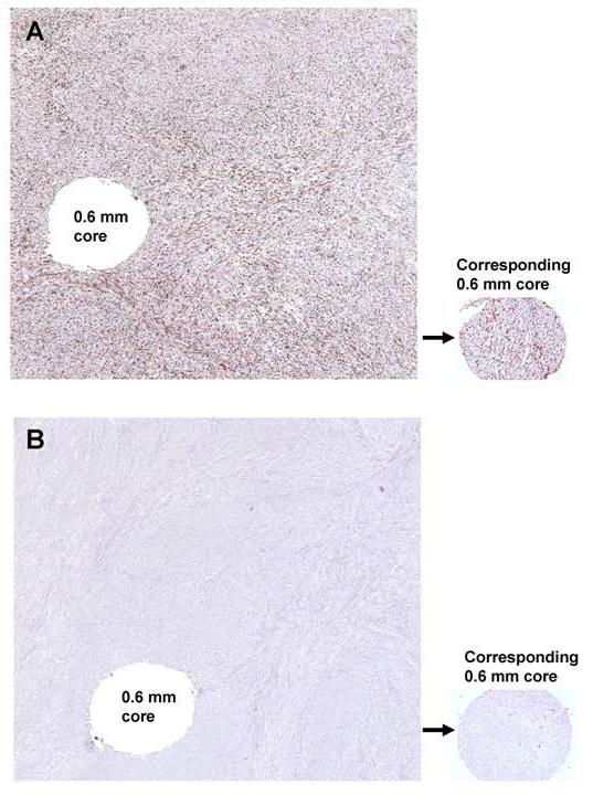
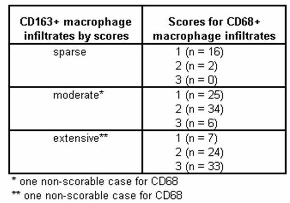
Web supplement Table 3: Comparison of the CD68 and CD163 immunostaining results in all LMS. Web supplement Figure 1: Whole tissue sections and corresponding 0.6 mm tissue cores showing LMS with A) case no.223: uniformly dense infiltrate of CD163 positive macrophages in the whole section and tissue core, B) case no.114: uniformly sparse infiltrate of CD163 positive macrophages in the whole section and tissue core.
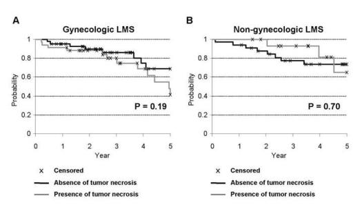
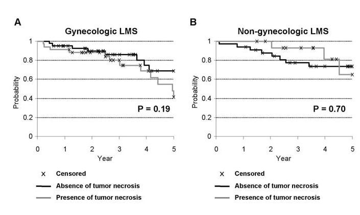
Web supplement Figure 2: Kaplan-Meier survival curves for the presence of tumor necrosis in (A) gynecologic and (B) non-gynecologic LMS.
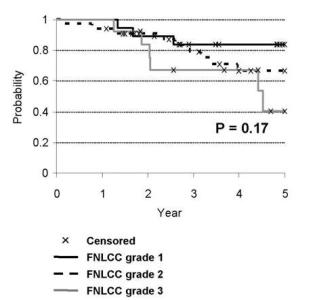
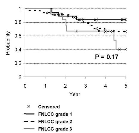
Web supplement Figure 3: Kaplan-Meier survival curves by FNLCC grade in non-gynecologic LMS.
|

